Buscar
-
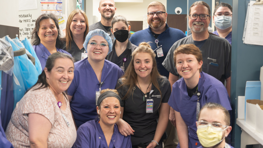
Departamento destacado: Patología
Celebrate Leap Day by leaping into the world of Pathology at Renown Health! Think of discovering a diagnosis like solving a mystery: the condition is the suspect, the nurses are the frontline police force and the doctors are the lieutenants or captains finalizing the results of the case. You may notice that one crucial role is missing on this list – the detectives. In the diverse network of healthcare, the detectives are a significant part of each patient’s mystery-solving care team and represent many roles across our health system. When it comes to figuring out the elaborate details of a growth, disease, organ abnormality or cause of death, one team of detectives, quite literally, goes as deep as possible. Those detectives are the team members within Renown Pathology. For each specialized field within medicine or surgery, the Pathology department is here to play a crucial role in accurate diagnoses. With each slide examined and each test meticulously conducted in their bright laboratories, these dedicated professionals shape a path towards wellness and recovery. Meet Your Anatomy Experts Whether you have a chronic disease that needs consistent testing, a high-risk birth that requires placenta testing, a suspected cancerous tumor that needs a biopsy or a gall stone that must be removed (or anything in between), Renown’s Pathology team steps in to provide biological answers to your body’s questions. This department offers the most comprehensive in-house diagnostic testing in the region, from routine histology to full pathology. As the busiest pathology department in northern Nevada, this team boasts the fastest turnaround times from respected experts, including: Pathologists Pathology Assistants Histotechnicians Histotechnologists Clinical Lab Assistants Let’s break down the complex nature of these team members' jobs by walking through their everyday responsibilities at work! Pathologists Pathologists are medical doctors who specialize in the study and diagnosis of disease. With every slide they scrutinize and every sample they analyze, pathologists unravel the mysteries of disease with precision and compassion. Their responsibilities include interpreting laboratory tests, analyzing tissue and fluid samples (obtained from a variety of different sources, including biopsies and surgeries), staging cancer diagnoses and providing diagnostic insights that guide treatment decisions. “Our job is to help the patients and their doctors figure out what’s wrong,” said Dr. Christie Elliott, Pathologist and Medical Director of the Clinical Laboratory at Renown Regional Medical Center. “As the bulk of our cases deal with cancer, almost every day we start with a tumor board alongside fellow surgeons, oncologists, radiologists and geneticists. From there, we order extra studies, run through our cases to make diagnoses, review slides and ensure all information goes into the charts, which is especially important as 70% of data in medical charts is from the lab. A patient’s history is everything.” Pathology Assistants With the steadiest of hands, pathology assistants, also known as PAs (not to be confused with physician assistants), guide the diagnostic journey from patient specimen to diagnosis. They can typically be found processing surgical and biopsy specimens (includes accessioning, gross examination, description, and sampling for microscopic analysis), preparing tissue samples for microscopic evaluation, helping the pathologist determine a cause of death for autopsies by conducting organ dissections and maintaining detailed records of all diagnostic findings. “As a PA, I still impact patient care without being directly patient-facing,” said Andrew Whitner, Pathology Assistant. “I handle 300-350 small tissue blocks a day. During dissections, I identify landmarks, document what I see and turn those landmarks into slides, looking for things that don’t look normal.” “Our job is 90% all about gross specimens, and we also do eviscerations for autopsies,” added Leslieann Haffner, Pathology Assistant. “We are trained on what normal looks like; our goal is to find the abnormal.” Histotechnicians Histotechnicians work behind the scenes to help transform ordinary tissue into extraordinary windows of insight, revealing the inner workings of the human body. As vital members of the Pathology team, histotechnicians embed tissue specimens in paraffin wax blocks (a process that preserves the tissue's structure for examination), cut thin sections of tissue from the paraffin blocks using a microtome, mount tissue onto glass slides and stain the tissue slides using histological stains to highlight structures or cells. “With all the patient specimens we work with, we get to see a lot of organs and learn what is causing the abnormalities,” said Reiny Hitchcock, Histotechnician. “I enjoy the opportunities to expand my knowledge, especially while working alongside the doctors.” “Our job can change by the week,” added Jessica Fahrion, Histotechnician. “One week I’ll be in the grossing room, and the next week I might be training in cytology." Histotechnologists In a world where every slide holds the key to a patient's future, histotechnologists are the champions of progress. One career ladder step above histotechnicians, these team members often have a broader scope of responsibilities, including more complex laboratory procedures, developing and validating new techniques, managing laboratory operations, interpreting results and troubleshooting technical issues. You can count on histotechnologists for validating antibodies and handling orders from pathologists, oncologists, emergency physicians and more. “My day always involves looking into cases, reading reports, getting orders together and working with pathologists to help them with their diagnoses; I also work a lot with immunohistochemistry, helping out with routine slides,” said Charles Koeritz, Histotechnologist. “I especially enjoy doing validations, which help maintain the integrity of lab testing and our diagnostic processes.” Clinical Lab Assistants Our pathology clinical lab assistants are the masters at “filling in the blanks,” assisting in whatever area needs it most, especially in cytology and the grossing room. They are essential aspects of the Pathology team, collecting and storing specimens for further testing, assisting in managing test results, gathering data, managing supply inventory and more. “As a Clinical Lab Assistant, I can be scheduled anywhere, from tissue cassetting to grossing,” said Ellie Somers, Clinical Lab Assistant. “Working in cytology is one of my favorite parts of my job. It’s very rewarding to work with the doctors to uncover what treatments will help each patient. We do cytology very well here.” The Bottom Line Even though the Pathology department doesn’t always experience a lot of patient face-to-face time, they interact with patients in a different way – by uncovering the story that is the inner workings of the human body, one slide and one sample at a time. “It’s important to remember that the slide IS a patient,” said Dr. Elliott. “We are constantly learning from every case so we can continue to provide the best patient care possible.” Take a Photo Tour of the Pathology Lab!
-

Optimizing Mammogram Screenings: A Genetic Approach to a Personalized Screening Schedule
Breast cancer screening has long been a cornerstone of women's healthcare. With 1 in 8 women diagnosed with breast cancer in their lifetime1, the United States Preventive Services Task Force (USPSTF) has developed screening recommendations to help detect early-stage cancer. Notably in 2023, the USPSTF revised the recommended age for biennial mammogram screenings for women with average risk to start at age 40 instead of 502, estimated to result in 19% more lives being saved3 by starting screening earlier. While initiating screening at an earlier age offers advantages to a wide demographic, concerns about the potential of over-screening prompted research into the feasibility of identifying women with lower breast cancer risk who could safely delay mammograms. While guidelines address high-risk individuals, a notable gap exists in providing recommendations tailored to those at lower risk. To gain insight into a patient's risk level, physicians are able to utilize genetic testing to understand an individual's genetic makeup, providing precise insights into their predisposition to various health conditions, including breast cancer. Armed with this genetic information, healthcare providers could craft tailored screening strategies that align with an individual’s specific risk profile. This genetic risk-based approach underscores the value of genetics in individualizing the onset of screening to help avoid over-screening and its associated costs. Surprisingly, genetic information is not currently being widely utilized to identify women at risk of breast cancer or other diseases in clinical practice, despite its potential to make a significant positive impact for patients. A recent retrospective analysis of 25,591 women from the Healthy Nevada Project4 sheds light on the potential benefits of this genetic risk-based approach. The study classified 2,338 (9.1%) of these women as having a low genetic risk for breast cancer. What's remarkable is that these women exhibited a significantly lower and later onset of breast cancer compared to their average or high-risk counterparts. This finding suggests that it might be safe for low-risk women to delay mammogram screening by 5 to 10 years without compromising their health.
-

What Is an Echo-Tech?
When it comes to our heart, keeping this vital source of life in tip-top shape is of utmost importance. Echo technologists or echocardiographers, otherwise known as "echo-techs," are charged with that mission, providing critical information that leads to life-saving interventions to keep our hearts beating strong. Adrianne Little, echocardiographer at Renown Health, breaks down the echo-tech's role in the health system, the educational path it takes to get there and the unique perks that come with the profession. What does an echo tech do? “Echo techs play a key role in the diagnosis and treatment of patients,” said Adrianne. “We are members of the cardiovascular imaging team that perform ‘heart ultrasounds’ or echocardiograms. Although we are most commonly known as echo techs, our official title is either ‘cardiac sonographer’ or ‘echocardiographer.’” Echo techs use imaging technology and sophisticated ultrasound equipment to produce images of the heart. These images show how well the heart functions, as well as the valves, chambers and blood flow. Echocardiograms are used to diagnosis and treat a variety of heart conditions such as murmurs, arrhythmias and heart failure. At the end of the day, the main goal of echo techs is to help our cardiovascular team provide the quickest and most accurate diagnoses to help with patient management and help them receive the highest standard of care. “When it comes to looking at the heart, we are part of the front-line team," said Adrianne. “We provide real time critical information that leads to life saving interventions down the road.”
-

Ladies! Get Screened for Breast Cancer
Early detection is a significant piece of the breast cancer puzzle. Susan Cox, Renown Health Director of Cancer Operations, discusses what you need to watch for and how the latest technology can help detect potential cancer sooner. When should women start getting breast exams? It depends on risk factors: Average-risk women: Most medical organizations recommend the first mammogram between 40 and 44. Higher-risk women: Dependent on their high risk, which will dictate when they start screening, but generally around the age of 30 and not before 25 years old.
-

Parkinson's Disease Know The Important Symptoms
Parkinson’s disease – you may have heard of it because Neil Diamond and Ozzy Osbourne were recently diagnosed with it. Or perhaps you know Michael J. Fox is a strong advocate and funds research through his foundation. Neurologist Jonathan Spivack, MD, discusses this disease, while physiatrist Stephanie Jones, DO, explains how physical therapy can help as a supplemental treatment. According to the Parkinson’s Foundation about ten million people worldwide currently have this disease. What is Parkinson’s Disease? “Parkinson’s disease is a neurodegenerative disease that progresses slowly and definitely, though at variable rates,” explains Dr. Spivack. “Symptoms go beyond the classic motor changes. It results from a loss of specific dopamine-producing brain cells. Specifically, this loss is likely due to a mix of genetic and environmental factors,” he adds. Dopamine allows communication between particular nerve cells responsible for movement. If you have Parkinson’s dopamine levels gradually drop, causing a loss of motor skills. Generally, most patients with the disease are over age 65. Early Signs and Symptoms Diagnosing Parkinson’s can be difficult as some of the symptoms happen during the natural aging process. The Parkinson’s Foundation identifies the following 10 early signs of PD: Tremors or shaking of your hand, fingers or chin Small handwriting Loss of smell Sudden movements during sleep Stiffness when walking or moving Constipation Softer or lower voice volume Mad facial expression Feeling dizzy or faint Hunching or stooping posture A single sign may not point to the disease, but if you (or a loved one) has multiple signs, talk to your healthcare provider.
Read More About Parkinson's Disease Know The Important Symptoms
-
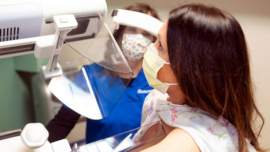
3D vs Whole Breast Ultrasound Which is Right for You
Breast cancer is the leading cause of cancer deaths in women in the U.S. That’s why early detection is so important. Dr. George Krakora, a radiologist with Renown Institute for Cancer, explains what to watch for and how new technology can lead to early detection. Most women know the importance of breast health and staying current with annual breast exams, but may not know that both screening guidelines and technology is evolving. So we asked George Krakora, MD, a radiologist for the Renown Institute for Cancer, what every woman should know about breast cancer detection and which screening method is right for them. First off, when should women start getting breast exams? Generally, women should start getting breast exams using mammography or ultrasound after they turn 40 years old. But we also want women ages 18 to 39 to talk to their primary care provider and ask for what’s called a formal risk assessment to see if screening is needed sooner. And you want to make sure your care provider is giving you a breast exam starting at age 25. It’s also a good idea to be familiar with how your breasts look and feel so you can report any changes to your care provider. What are the risk factors for breast cancer? Are there any preventive steps women can take? There a few risk factors you can’t control, like your age, family history of breast or other cancers, and if you have dense breast tissue. Your risk for breast cancer increases as you get older, and most breast cancers are diagnosed after age 50. Knowing your family history is important because a history of cancer and shared lifestyle can raise your risk. Your breast density can also increase your risk: Women with high breast density are four-to-five times more likely to get breast cancer than women with low breast density. But the good news is there are quite a few things you can do to prevent breast cancer, like not smoking, watching your alcohol intake, and maintaining a healthy weight with good diet and exercise. There are a lot of newer screenings out today. What is the difference between 2-D and 3-D mammography? In a 2-D mammogram, the tech takes X-rays of the breast. These pictures can show the radiologist if there are any lumps or tumors you might not be able to feel. In 3-D mammography, the process is largely the same but more X-rays are taken and it takes a few seconds longer for each image. This kind of exam detects 41 percent more cancers and reduces the number of false-positive results given to patients. This improvement in technology is great for both patients and their care providers. 3-D mammography provides better images of the breast, which allow doctors to more clearly diagnose and avoid false positives, especially in women with dense breast tissue. And what about a whole breast ultrasound. What is that? A whole breast ultrasound uses sound waves to detect cancerous tumors in the breast without using any radiation — it’s an ultrasound just like pregnant women get to check up on their baby. And the exam only takes about 20 minutes. We recommend these exams for patients whose mammograms have shown that they have dense breast tissue. Dense breast tissue can make it harder for doctors to see any abnormalities, lumps or tumors in a mammogram, so this technology ensures better early detection.
Read More About 3D vs Whole Breast Ultrasound Which is Right for You
-

The Facts About Menopause and Early Menopause
Menopause is something that every woman experiences at some point in her lifetime. Learn what to expect and how you can help manage the symptoms and health risks. Most women don’t experience menopause until their 50s, but certain factors such as chromosomal abnormalities, glandular problems and chemotherapy can cause early menopause before the age of 40. No matter what your age, it’s a good idea be aware of the risks and treatments available to maintain a comfortable and healthy lifestyle. Health Risks of Menopause Two of the biggest health risks posed to women who have gone through menopause are bone density loss and risk of cardiovascular disease. Bone loss can be treated with bisphosphonate and estrogens. “Calcium with vitamin D and weight bearing exercise will also limit bone loss,” says Vickie Tippett, MD and OB/GYN at Renown Health. For cardiovascular risk, a healthy lifestyle is key. Discontinuing tobacco use, getting regular exercise and maintaining a healthy weight and diet all help reduce a woman’s risk of cardiovascular disease. Managing Discomforts of Menopause One of the most common complaints about menopause is the discomfort of hot flashes. “Hot flashes can be treated with systemic estrogen alone or in combination with progesterone or another agent similar to estrogen,” Dr. Tippett says. “Non-hormonal medications such as SSRIs and antidepressants also work.” Vaginal dryness, another common symptom of menopause, can also be treated with estrogen, estrogen-like compounds and personal lubricants. Pills, patches, creams and many other formulations are available to help alleviate discomfort. Knowing when, why and what to expect when it comes to menopause can help make the transition easier. Learn the facts about menopause in the infographic below.
Read More About The Facts About Menopause and Early Menopause
-

What Every Woman Needs to Know About Dense Breast Tissue
In honor of International Women’s Day, we’re working to spread the word about taking care of your breast health and encouraging the women in your life to do the same. Heather Reimer is on a mission — a mission to educate women everywhere about breast tissue type. For women with dense breasts, knowing your breast tissue type is absolutely critical, as cancers embedded in dense breast tissue are not always detectable with a mammogram alone. Dense breast tissue requires a breast ultrasound screening to get a complete breast health picture. Whole Breast Ultrasound for Dense Breast Tissue Heather knows this firsthand. She has dense breasts, and in this video she shares her story about finding breast cancer during a breast ultrasound screening — cancer that went undetected with her mammogram screening alone. As a result of that experience, Heather founded Each One. Tell One. — a movement to encourage women to pass along this information to others and to prompt those with dense breast and implants to consult with their doctor to schedule a whole breast ultrasound screening. To schedule a mammogram or a whole breast ultrasound, call 775-982-8100.
Read More About What Every Woman Needs to Know About Dense Breast Tissue